Overview
Rectal cancer is one of the most common cancers you will see on a lower-GI posting, and it is also one where radiotherapy plays a central role. The disease is usually managed by a multidisciplinary team (MDT), and treatment is decided by the stage of the cancer, the closeness to surgical margins and whether organ preservation (sphincter preservation) is a goal.
Rectal cancer differs from colon cancer because of its location in the narrow, fixed space of the pelvis. Because of its location, local pelvic recurrence is a real risk, and this is exactly the problem that radiotherapy helps to solve.
Anatomy
The rectum is the last part of the large bowel, about 15 cm in length measured from the anal verge at colonoscopy or sigmoidoscopy. We can divide the rectum into three parts:
- Lower third — 0–5 cm from the anal verge.
- Middle third — 5–10 cm from the anal verge.
- Upper third — 10–15 cm from the anal verge.
This matters because lower tumours are closer to the anal sphincter and are more likely to need radiotherapy and to threaten the chance of saving the sphincter.
Lymphatic drainage
- Mesorectal nodes
- Presacral nodes
- Internal iliac nodes
- Obturator nodes
Cross-sectional anatomy
The wall of the rectum has layers—from the inside out: mucosa, submucosa, muscularis propria, then the surrounding fat.
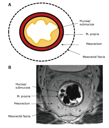
The mesorectum and the mesorectal fascia
The rectum is wrapped in a layer of fatty tissue called the mesorectum, which contains the blood vessels and the local lymph nodes. The mesorectum is enclosed by a thin envelope called the mesorectal fascia. This fascia is very important for two reasons:
- Surgery: the standard operation, total mesorectal excision (TME), removes the rectum together with the whole mesorectum by dissecting along this fascia. Removing it as an intact package greatly lowers the chance of local recurrence.
- Imaging: on MRI the fascia becomes the circumferential resection margin (CRM). If the tumour reaches or threatens the fascia, the CRM is “threatened”, and these patients usually need radiotherapy before surgery.
Diagnosis and Staging Work-up
Before any treatment is planned, every patient needs a standard set of investigations so the MDT knows exactly what they are dealing with. The basics you should know are:
- Digital rectal examination (DRE) — a simple finger examination that tells you how far the tumour is from the anal verge and whether it is mobile or fixed.
- Colonoscopy confirms the diagnosis under the microscope and checks the rest of the colon for other tumours or polyps.
- Biopsy: the most common histology is adenocarcinoma.
- MRI of the pelvis — the key local staging test. It shows how deep the tumour is (T), suspicious nodes (N), and whether the mesorectal fascia or CRM is threatened, which can result in a positive surgical margin.
- CT of the chest, abdomen and pelvis — looks for spread to other organs, most commonly the liver and lungs (M).
- Carcinoembryonic antigen (CEA) — a blood tumour marker used as a baseline and to help with follow-up.
Staging: TNM Basics
Staging uses the TNM system: T for how deep the tumour has grown, N for lymph nodes, and M for spread to distant organs.
| Category | What it means |
|---|---|
| T1 | Tumour invades the submucosa, just under the lining. |
| T2 | Tumour invades the muscularis propria, the muscle layer. |
| T3 | Tumour grows through the muscle into the surrounding fat. |
| T4 | Tumour invades the peritoneum or a nearby organ. |
| N0 / N1 / N2 | No nodes / 1–3 involved nodes / 4 or more involved nodes. |
| M0 / M1 | No distant spread / distant spread present, for example liver or lung. |
Treatment Overview
Treatment is always decided by the MDT, which includes surgeons, oncologists, radiologists and pathologists. The three main tools are surgery, radiotherapy and chemotherapy, used in different combinations depending on the stage.
Surgery
Surgery is the main curative treatment for most rectal cancers. The standard operation is total mesorectal excision (TME), removing the rectum and its mesorectum as one intact package. Depending on tumour location, the surgeon may be able to rejoin the bowel and spare the sphincter (anterior resection) or may need to remove the anus and form a permanent stoma (abdominoperineal resection). Very small, early tumours can occasionally be excised locally through the anus.
Role of radiotherapy
Radiotherapy is mainly used before surgery (neoadjuvant) for locally advanced tumours—those that are T3–T4, node-positive, or close to the mesorectal fascia. Treating before surgery shrinks the tumour, makes complete removal more likely, and lowers the risk of the cancer coming back in the pelvis.
Early tumours (T1–T2 N0) with a clear margin on MRI usually do not need radiotherapy and go straight to surgery.
Chemotherapy
Chemotherapy is used in two ways: a low dose given at the same time as radiotherapy (concurrent) as a sensitizer, and full-dose chemotherapy given before or after surgery to treat any microscopic spread. The most common drug given alongside radiotherapy is capecitabine (an oral tablet), or sometimes 5-fluorouracil (5FU) as an IV infusion.
Sequencing of multimodality therapy
Chemo-radiotherapy → surgery → chemotherapy
Chemotherapy → chemo-radiotherapy → surgery
Chemo-radiotherapy → chemotherapy → surgery
Surgery → chemotherapy → chemoradiotherapy → chemotherapy
Radiotherapy in Rectal Cancer
Radiotherapy dose is 45 Gy in 25 fractions to the primary rectal tumour, the grossly enlarged nodes, the mesorectum and lymph-node stations including the presacral, obturator and internal iliac nodes. A boost of 5.4 Gy in 3 fractions is given to the primary tumour and grossly enlarged nodes, making the total dose 50.4 Gy in 28 fractions. Alternatively, a simultaneous integrated boost (SIB) technique can be used in 25 fractions, where the boost (50 Gy, 2 Gy per fraction) is delivered simultaneously.
How a radiotherapy course is planned
- Simulation: a planning CT scan is taken in the treatment position, usually supine with a comfortably full bladder (>250 ml), to push small bowel out of the radiation field.
- A simulation MRI without contrast is also performed and fused with the planning CT scan.
- Diagnostic pelvic MRI and PET, if available, are also fused with the planning CT scan.
- Contouring:
- Primary tumour (GTVp)
- Grossly enlarged nodes (GTVn)
- Elective clinical target volumes including the mesorectum and nodal stations such as presacral, internal iliac and obturator nodes
- Organs at risk: small bowel or peritoneal space, bladder, bone marrow, femoral heads and proximal femur
- Radiotherapy delivery technique: intensity-modulated radiotherapy (IMRT) using RapidArc (VMAT).
References and further reading
- National Cancer Institute: Rectal Cancer Treatment (PDQ®), Health Professional Version — evidence summaries and supporting literature on staging and treatment.
Further-reading link checked on 5 September 2026. The editorial update date records changes to this page’s presentation and references; it is not a clinical evidence-review date.